From 25.- 27.10.2021 the Research Unit organised an Autumn Workshop on Image Segmentation, Analysis and Bio-Mechanical Modeling at Heidelberg University. Here the participants learned, among other things, to create images and videos in various programmes as featured in this article.

They learned the basics and advanced features of PlantSeg, ilastik, and MorphoMechanX with additional plugins to analyse plant organ development. The workshop was led by the FOR members Gabriella Mosca, Adrian Wolny and Sören Strauss.
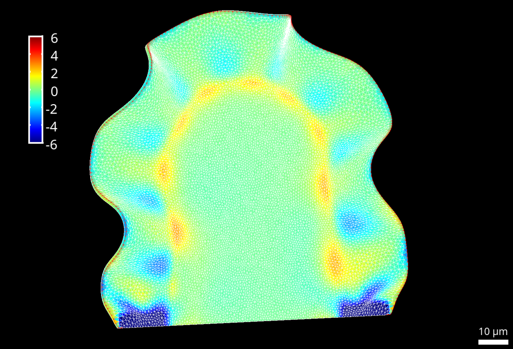
(Video on the left: Relative volume increase of cells in an idealised section of Arabidopsis embryo due to growth (so pressurized grown template / pressurized non-grown template). Source Bassel et al. 2014, PNAS. All the simulations were performed in MorphoMechanX (www.morphographx.org/morphomechanx) and use the Finite Element Method to solve continuum mechanics problems)
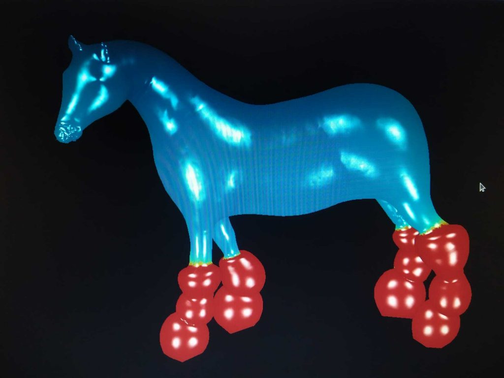
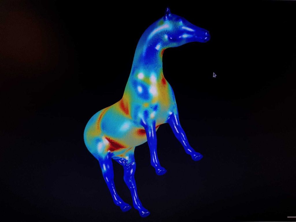
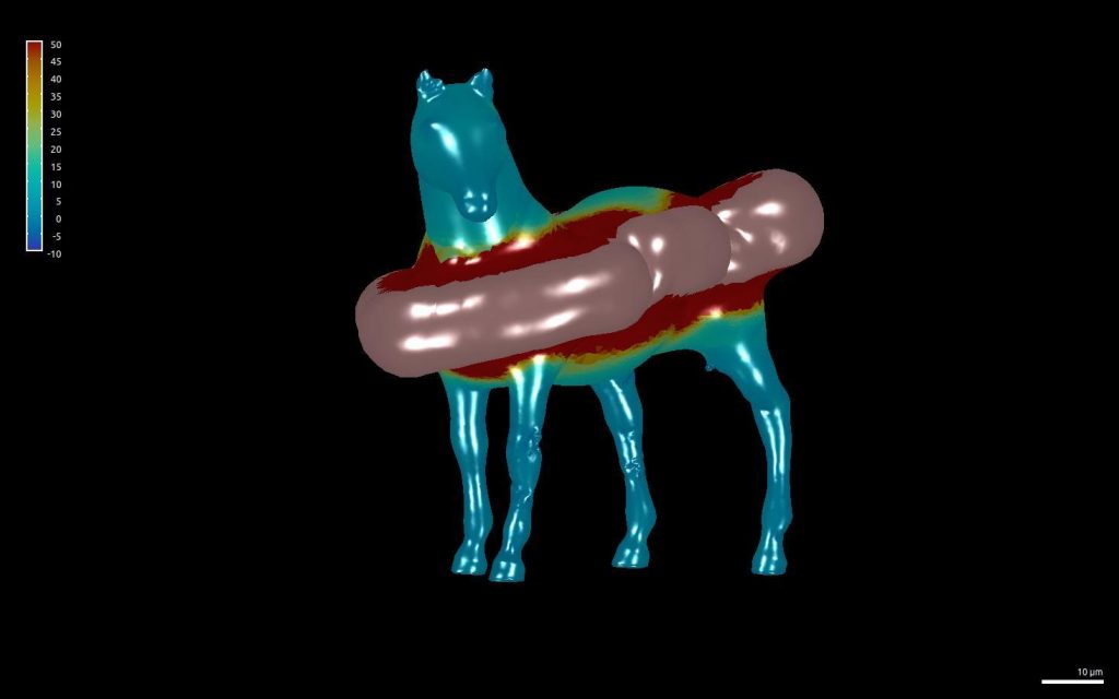
During the workshop, the participants were also allowed to have some fun with the programs. So did they inflate a horse template made of membrane elements. The mesh comes from a mesh database, and the heatmap is still trace of cauchy stress. While most of these experiments were physically not correct, they contributed to a further understanding of the program.


